Contribute
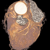
| Technology - Multidetector CT Angiography: A Breakthrough In The Detection Of Coronary Artery Disease |
Thomas J. Brady, MD
09/05/2006
According to the World Health Organization, coronary artery disease
(CAD) is one of the leading causes of death worldwide, accounting for
17 million deaths per year. Although it was once considered a “western
disease,†today more than 60% of cases occur in developing countries.
While a number of non-invasive diagnostic tests, such as nuclear
scanning and echocardiography, provide valuable information about the
heart and coronary arteries, the gold standard for the detection and
evaluation of CAD is coronary angiography, which is performed nearly
1.5 million times a year in the U.S. Despite its undisputed clinical
value, coronary angiography is costly, invasive, and poses some risks,
therefore it is indicated only for patients determined to be at high
risk for CAD.
A NON-INVASIVE ALTERNATIVE
Because early detection and treatment of CAD can significantly reduce
morbidity and mortality, there has been an ongoing, widespread interest
in finding a non-invasive alternative to coronary angiography that is
equal to, if not better than, the current gold standard. The modality
showing the greatest promise is cardiac computed tomography (CT).
When the first CT scanners were introduced into clinical practice in
the 1970s, cardiac imaging was not feasible, as the time required to
acquire an image—approximately five minutes—ruled out a motionfree
image of the heart and coronary vessels.
Over the ensuing
decades, CT technology has improved dramatically. High spatial and
temporal resolution—which are required for clinically useful cardiac
imaging—have been achieved with the introduction of multiple rows of
detectors (from 4 to 16 and, most recently, 64), faster gantry
rotation, and sophisticated ECG cardiac-gating techniques.
Massachusetts General Hospital recently became the first hospital in
New England to acquire and begin using a 64-slice, multi-detector CT
(MDCT) scanner, which is used exclusively for the evaluation of cardiac
patients. This state-of-the-art technology virtually freezes the
heart’s detail and clarity. This is achieved in a single breathhold
with a scan time of just 8-12 seconds, a radiation exposure equivalent
to a chest CT scan, and a door-to-door time of approximately 15 minutes.
INDICATIONS
Presently, the indications for the use of Mass General’s 64-slice cardiac CT scanner are to:
• rule out significant CAD (e.g., as part of a routine pre-operative exam) in patients with low and regular heart rates
• rule out CAD in patients with atypical chest pain and an intermediate risk of CAD
• evaluate patients with inconclusive ECG stress tests
• visualize anomalous coronary arteries
• establish the patency of bypass grafts
• visualize the cardiac anatomy for congenital malformations, pulmonary venous return, and masses
• detect and quantify coronary plaque
EXCLUSION CRITERIA
• arrhythmias
• pregnancy
• impaired renal function
• allergy to the iodinated contrast agent
• myeloma
Although not yet established through clinical trials, it is anticipated
that as a result of earlier diagnosis and treatment, cardiac CT will
reduce morbidity and mortality among patients with suspected CAD.
A ROLE IN PATIENT MANAGEMENT
Current evidence suggests that cardiac CT may also have an important
role in the management of patients with established CAD. It is the only
non-invasive technique in widespread clinical use that can obtain
information about the amount and composition of plaques in the coronary
arteries, which are the cause of most acute coronary events. Thus,
cardiac CT could become a valuable tool for evaluating patients who
have had a mild myocardial infarction to determine whether they are
candidates for aggressive treatment aimed at reducing their risk of a
subsequent coronary event or sudden cardiac death.
Investigators in the Massachusetts General Hospital Department of
Radiology and Division of Cardiology have been actively involved in
cardiac CT research during the past five years, and have published
numerous papers on their work (see Selected References). Research
currently under way by this group is focused on a variety of new
potential applications for cardiac CT. These include functional studies
of myocardial perfusion and the role this technology may play in
triaging ED patients with acute chest pain.
Undoubtedly the
applications of cardiac CT will increase as investigators at Mass
General and elsewhere continue to study its potential and as the
technology continues to improve. But there is no question that this
state-of-the-art technology is already having a major impact on patient
care.
SELECTED REFERENCES
Hoffman U, Moselewski F, Cury R, Ferencik M, Jang I, Diaz L, Abbara S,
Brady T, Achenbach S. Predictive value of 16-slice multi-detector
spiral CT to detect significant obstructive coronary artery disease in
patients at high risk for CAD: patient vs. segment-based analysis.
Circulation 2004; 110:2638-2643.
Achenbach S, Moselewski F,
Ropers D, Ferencik M, Hoffman U, MacNeill B, Pohle K, Baum U, Anders K,
Jang I, Daniel W, Brady T. Detection of calcified and non-calcified
coronary atherosclerotic plaque by contrast-enhanced, submillimeter
multidetector spiral CT:
a segmentbased comparison to IVUS. Circulation 2004;109:14-17
You may also access this article through our web-site http://www.lokvani.com/